전립선 자료실
페이지 정보
본문
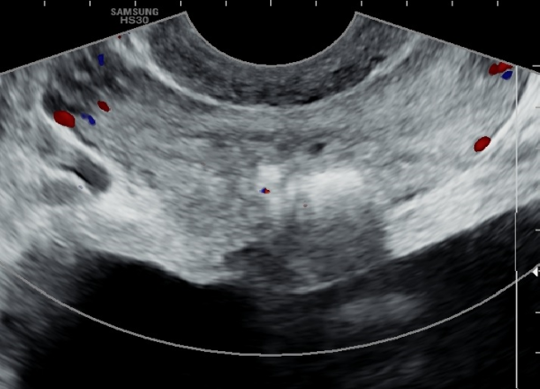
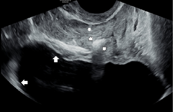
6년 동안 회음부의 통증과 배뇨장애로 타의원에서 약물만 복용을 했으나 증상의 호전이 없다고 내원 하신 분의 내원 당일
검사한 경직장 전립선 초음파 사진입니다.
Transrectal prostate ultrasound image taken on the day of visit from a patient who had been experiencing perineal pain and urinary dysfunction for six years and had only been on medication at another clinic without symptom improvement.
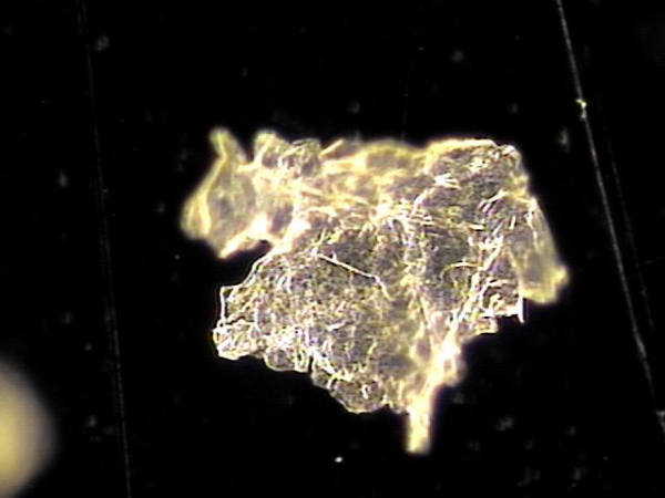
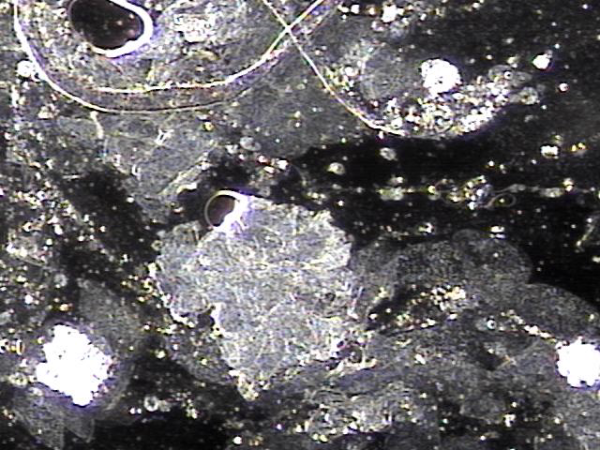
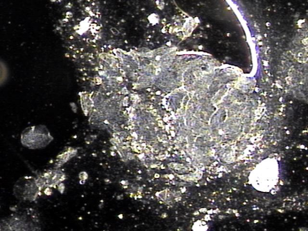
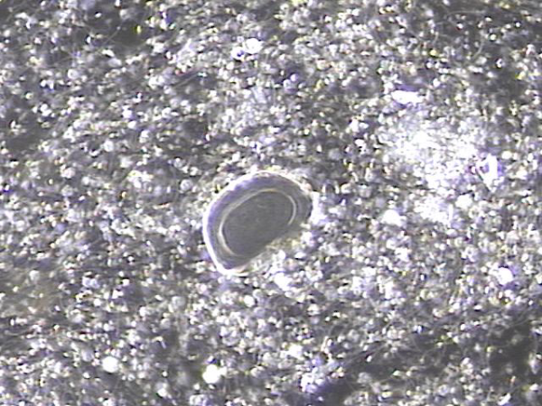
주 2회 전립선의 표적 치료중 전립선과 정관과 정낭 그리고 사정관에 쌓여 있는 탈락된 상피 세포가 치료후 관찰된 현미경학적 검사 자료 입니다.
Microscopic examination findings observed after targeted prostate treatment twice a week,
showing exfoliated epithelial cells accumulated in the prostate, vas deferens, seminal vesicles, and ejaculatory ducts.
주 2회 전립선의 표적 치료중 전립선과 정관과 정낭 그리고 사정관에 쌓여 있는 탈락된 상피 세포가 치료후 관찰된 현미경학적 검사 자료 입니다.
Microscopic examination findings observed after targeted prostate treatment twice a week,
showing exfoliated epithelial cells accumulated in the prostate, vas deferens, seminal vesicles, and ejaculatory ducts.
주 2회 전립선의 표적 치료중 전립선과 정관과 정낭 그리고 사정관에 쌓여 있는 탈락된 상피 세포가 치료후 관찰된 현미경학적 검사 자료 입니다.
Microscopic examination findings observed after targeted prostate treatment twice a week,
showing exfoliated epithelial cells accumulated in the prostate, vas deferens, seminal vesicles, and ejaculatory ducts.
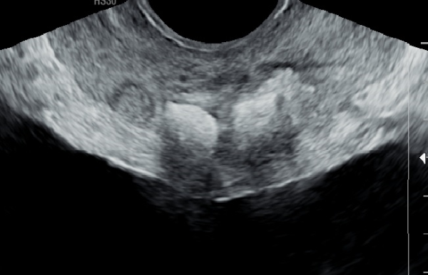
4개월동안 주 2회 전립선의 표적 치료후 사정관 입구와 전립선에 고여 있던 결석이 감소 하고 있는 전립선의 경직장 초음파 추적 사진입니다.
Transrectal ultrasound follow-up images of the prostate after four months of targeted prostate treatment twice a week, showing a reduction in the stones that had accumulated at the entrance of the ejaculatory ducts and within the prostat
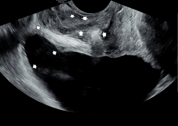
첫 진료 당일 검사한 경직장 전립선의 초음파 검사상 정낭의 낭종이 관찰되는 자료입니다.
Transrectal ultrasound examination on the first visit showing the presence of seminal vesicle cysts.
주 2회 전립선의 표적 치료중 전립선과 정관과 정낭 그리고 사정관에 쌓여 있는 탈락된 상피 세포가 치료후 관찰된 현미경학적 검사 자료 입니다.
Microscopic examination findings observed after targeted prostate treatment twice a week,
showing exfoliated epithelial cells accumulated in the prostate, vas deferens, seminal vesicles, and ejaculatory ducts.
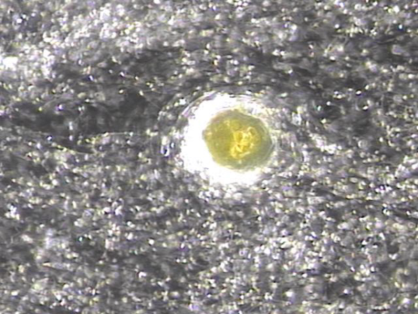
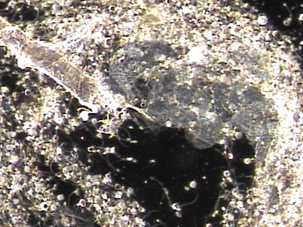
주2회 전립선의 표적 치료후 배출된 전립선 결석과 정낭속에 있는 정자들의 현미경 학적 사진입니다.
Microscopic images of expelled prostatic calculi and sperm within the seminal vesicles after twice-weekly targeted prostate therapy.
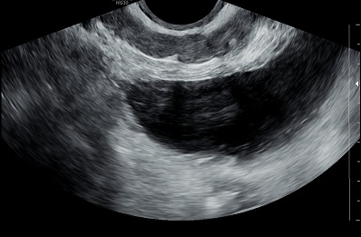
주 2회 4개월동안 전립선의 표적 치료후 정낭의 낭종들이 치료된 추적 경직장 전립선 초음파 사진입니다.
Follow-up transrectal ultrasound images of the prostate showing treated seminal vesicle cysts after four months of twice-weekly targeted prostate therapy.
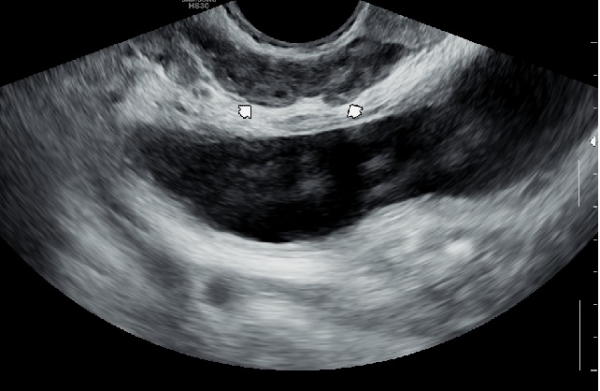
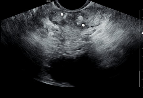
내원 당일 검사 한 경직장 전립선의 초음파 측면 사진상 사정관 입구의 순환 장애로 사정관이 확장된 사진 자료입니다.
A lateral transrectal ultrasound image of the prostate taken on the day of the visit, showing dilation of the ejaculatory duct due to circulatory dysfunction at the ejaculatory duct opening.
주 2회 전립선의 표적 치료중 전립선과 정관과 정낭 그리고 사정관에 쌓여 있는 탈락된 상피 세포가 치료후 관찰된 현미경학적 검사 자료 입니다.
Microscopic examination findings observed after targeted prostate treatment twice a week,
showing exfoliated epithelial cells accumulated in the prostate, vas deferens, seminal vesicles, and ejaculatory ducts.
주 2회 전립선의 표적 치료후 사정관 입구의 결석이 부분적으로 감소하고 사정관에 쌓인 탈락된 상피 세포가 치료되면서 확장된 사정관이 없어진
경직장 전립선 추적 검사 자료입니다.
A follow-up transrectal ultrasound examination of the prostate after twice-weekly targeted prostate treatment, showing partial reduction of stones at the ejaculatory duct opening and resolution of the dilated ejaculatory duct as the accumulated desquamated epithelial cells in the ejaculatory duct were treated.
주2회 전립선의 표적 치료후 배출된 전립선 결석의 현미경학적 사진입니다.
Microscopic images of expelled prostatic calculi after twice-weekly targeted prostate therapy.
요의를 참지 못하고 화장실에 도착하기 전 소변이 새어 나오는 절박성 요실금의 원인중 치료해야할 항문주위 결석들
Perianal calculi that need to be treated as one of the causes of urge urinary incontinence,
where urine leaks before reaching the toilet due to an inability to hold the urge to urinate
- 다음글전립선자료실 25.03.13
댓글목록
등록된 댓글이 없습니다.


