전립선자료실
페이지 정보
본문
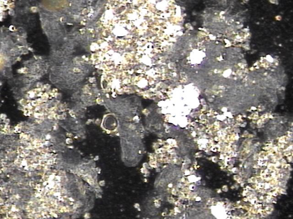
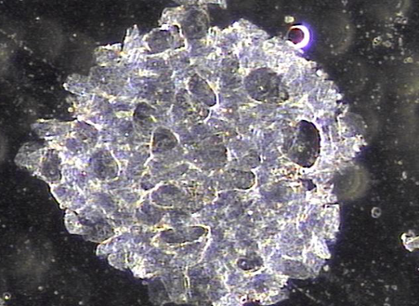
주2회 전립선의 표적 치료후 치료된 상피 세포 덩어리의 현미경학적 자료입니다.
"This is the microscopic data of the epithelial cell clusters treated after targeted prostate therapy twice a week."
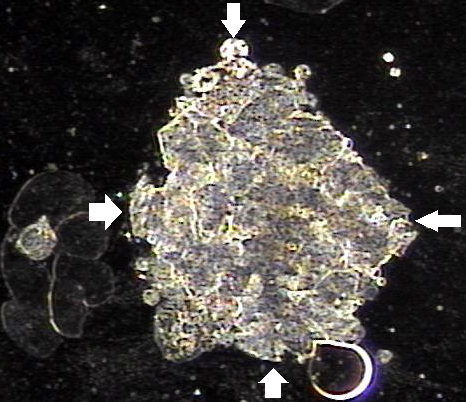
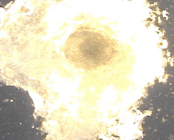
주2회 전립선의 표적 치료후 치료된 상피 세포 덩어리의 현미경학적 자료입니다.
"This is the microscopic data of the epithelial cell clusters treated after targeted prostate therapy twice a week."
개인의 차가 있을수 있으나
평균적으로 항문에서 전립선까지 거리는 6Cm,
정낭까지의 거리는10Cm, 그리고
정낭의 크기는 가로5Cm,세로2.5Cm,두께1Cm 즉 항문에서 정낭 첨부까진 15Cm 그리고
정관은 좌우 각각 45Cm 항문에서 정관까지 6Cm 이상
사정관까지도 항문에서 6Cm이상 접하여
전립선의 표적치료를 해야하며
사람마다 둔부를 구성하는 대둔근과 중둔근, 소둔근의 크기가 달라
표적치료시 서로 노력하여
만성 전립선염과 전립선 비대증, 전립선의 낭종과 전립선의 결석과 전립선의 암 그리고
정낭의 낭종 고환의 미석증과 만성골반통 증후군등의 반드시
치료되는 전립선의 표적치료를 하러가야합니다.
이따가 뵙겠습니다.
서울가정의학과의원 드림.
While there may be individual variations, on average:
- The distance from the anus to the prostate is 6 cm.
- The distance to the seminal vesicles is 10 cm.
- The size of the seminal vesicles is 5 cm in width, 2.5 cm in height, and 1 cm in thickness, meaning the distance from the anus to the tip of the seminal vesicle is approximately 15 cm.
- The vas deferens extends 45 cm on each side, with at least 6 cm from the anus to the vas deferens and the ejaculatory ducts.
The Importance of Targeted Prostate Treatment
Due to this anatomical structure, targeted prostate treatment is essential. Since the size of the gluteal muscles (gluteus maximus, medius, and minimus) varies among individuals, precise efforts are needed during treatment.
This treatment is crucial for effectively managing and resolving conditions such as:
- Chronic prostatitis
- Benign prostatic hyperplasia (BPH)
- Prostatic cysts and calcifications
- Prostate cancer
- Seminal vesicle cysts
- Testicular microlithiasis
- Chronic pelvic pain syndrome (CPPS)
We highly recommend undergoing targeted prostate treatment to address these issues.
See you soon.
Best regards,
Seoul Family Medicine Clinic
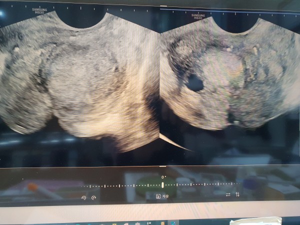
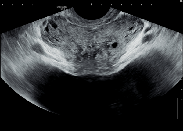
처음 본 의원애 내원하여 경직장 전립선 초음파 검사상 전립선의 다발성 낭종이 관찰된 사진
"A patient visited the clinic for the first time, and multiple prostate cysts were observed on the transrectal prostate ultrasound scan."
주2회 전립선의 표적 치료후 치료된 상피 세포 덩어리의 현미경학적 자료입니다.
"This is the microscopic data of the epithelial cell clusters treated after targeted prostate therapy twice a week."
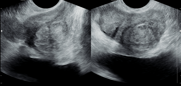
주 2회 전립선의 표적 치료후 다발성 낭종이 치료되고 있는 경직장 전립선 초음파 사진
"A transrectal prostate ultrasound image showing the treatment of multiple cysts after targeted prostate therapy twice a week."
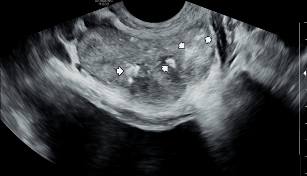
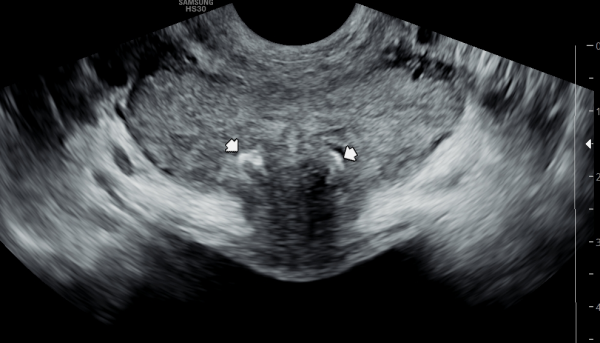
서울가정의학과의원에 내원 당일 경직장 전립선 초음파 검사상 양쪽 사정관 입구의 결석과 좌측 전립선의 중심 구역에 생긴 결석이 관찰된 자료입니다.
"This is the data from the day of the visit to the Seoul Family Medicine Clinic, showing the presence of stones at the entrances of both ejaculatory ducts and a stone located in the central zone of the left prostate, as observed on the transrectal prostate ultrasound."
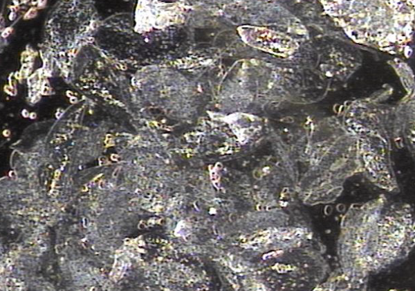
주2회 전립선의 표적 치료후 치료된 상피 세포 덩어리의 현미경학적 자료입니다.
"This is the microscopic data of the epithelial cell clusters treated after targeted prostate therapy twice a week."
주2회 전립선의 표적치료후 양측 사정관 입구의 결석과 좌측 전립선의 중심 구역에 생긴 결석이 치료 되고 있는 경직장 전립선 초음파 사진
"This is a transrectal prostate ultrasound image showing the treatment of stones at the entrances of both ejaculatory ducts and a stone in the central zone of the left prostate, following targeted prostate therapy twice a week."
댓글목록
등록된 댓글이 없습니다.


