전립선자료실
페이지 정보
본문
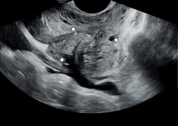
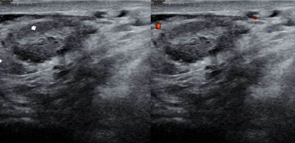
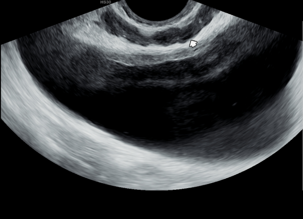
수년전부터 회음부 통증과 배뇨장애로 내원 당일 경직장 측면 전립선 초음파 사진상 사정관 입구의 결석과 전립선내 낭종과 커진 결절을 보이는 초음파 사진입니다.
A lateral transrectal prostate ultrasound image taken on the day of the visit due to years of perineal pain and voiding dysfunction, showing a calcification at the ejaculatory duct opening, a cyst within the prostate, and an enlarged nodule.
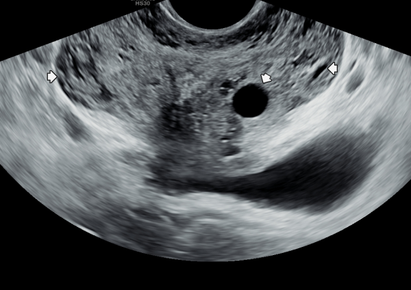
처음 본 의원애 내원하여 경직장 전립선 초음파 검사상 전립선의 다발성 혈낭종이 관찰된 사진
"A patient visited the clinic for the first time, and multiple prostate hemorragic cysts were observed on the transrectal prostate ultrasound scan."
특이한 사항은 6년전 심근 경색의 위험한 순간, 심장에 관상동맥확장술 즉 shunt operation for dilation for coronary artery를 시행후 지속적인 항혈소판제의
약을 복용후 전립선내 다발성 혈낭종이 생긴 사례입니다.
A notable case involves the development of multiple hemorrhagic cysts in the prostate following continuous use of antiplatelet medication after undergoing a coronary artery dilation procedure (shunt operation for dilation of the coronary artery) during a critical moment of myocardial infarction six years ago.
사정관 입구의 결석으로 정낭과 정관의 순환 장애로 생긴 정낭의 다발성 낭종의 경직장 전립선의 초음파 사진입니다.
This is a transrectal prostate ultrasound image showing multiple seminal vesicle cysts caused by circulatory obstruction in the seminal vesicles and vas deferens due to a stone at the ejaculatory duct opening.
첫 내원 당일 경직장 전립선 초음파 사진의 측면 사진과 정면 사진상 수년간 복용중인 혈전용해제 복용으로 다발성 혈낭종이 관찰되는 초음파 사진입니다.
This transrectal prostate ultrasound image, taken on the patient's first visit, shows both lateral and frontal views. Multiple hemorrhagic cysts are clearly visible, which are suspected to be associated with the long-term use of thrombolytic (blood-thinning) medication. The findings are concerning and warrant careful monitoring and appropriate medical intervention.
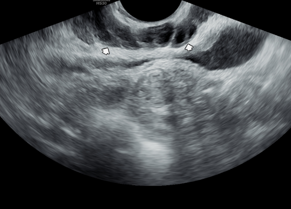
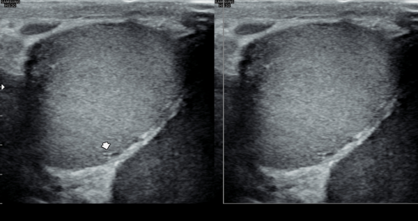
처음 내원 당일 고환의 초음파 검사상 정관의 순환 장애로 고환의 섬유화가 관찰되는 초음파 사진입니다.
This is a scrotal ultrasound image taken on the patient's first visit, showing testicular fibrosis due to impaired circulation in the vas deferens.
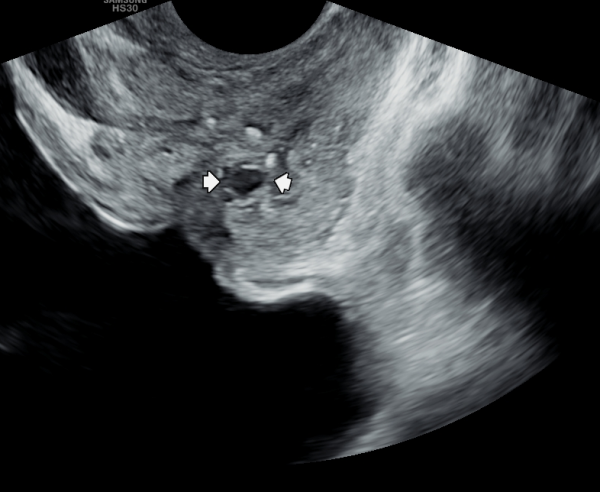
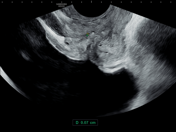
주 2회 전립선의 표적 치료후 전립선의 결절 크기가 감소하고 혈낭종이 치료 되면서 숨겨져 있던 사정관 입구의 결석과
전립선내 결석이 관찰되는 초음파 사진입니다.
This ultrasound image shows that after twice-weekly targeted prostate treatments, the size of the prostatic nodule has decreased and the hemorrhagic cysts have been treated, revealing previously hidden stones at the ejaculatory duct openings and within the prostate.
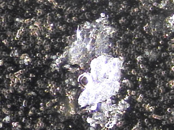
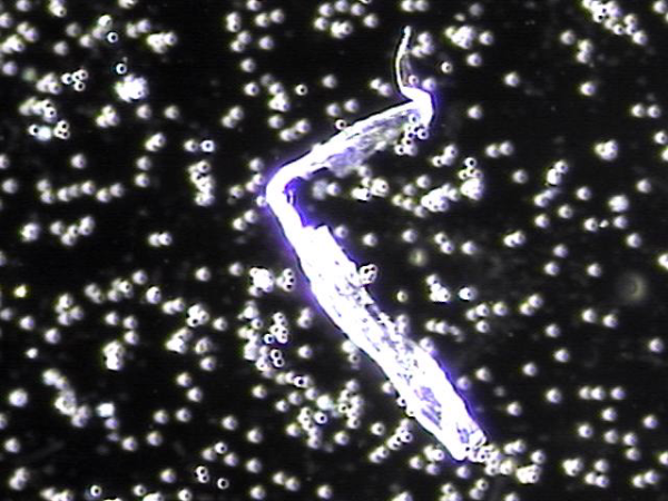
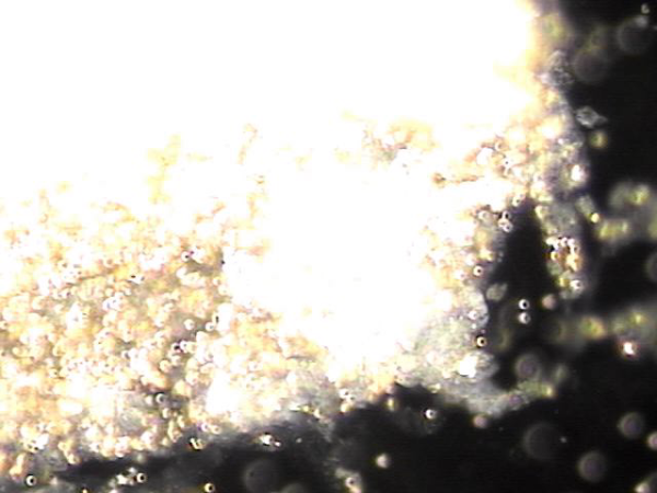
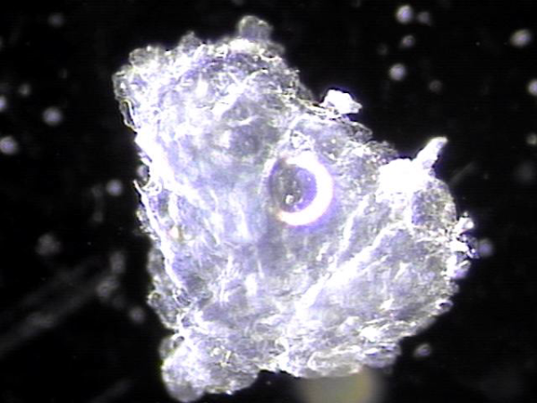
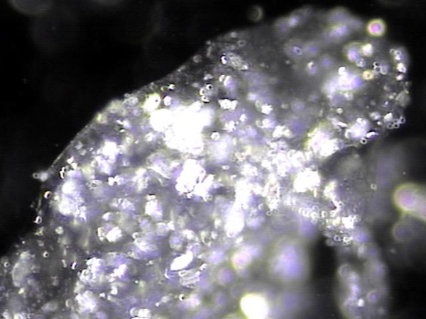
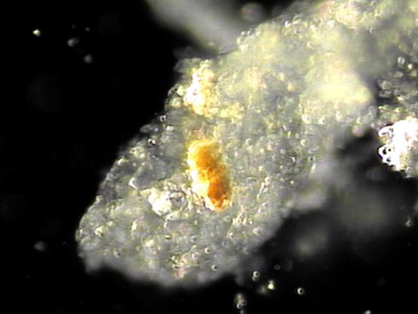
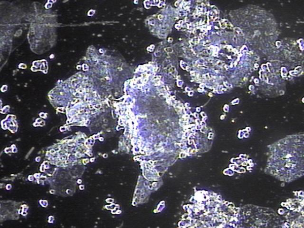
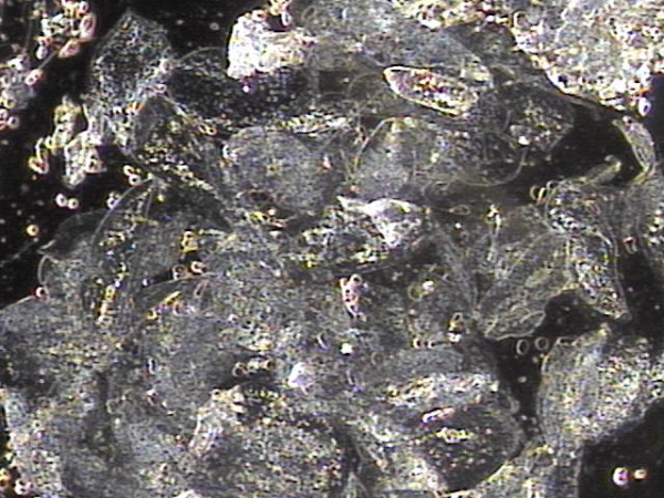
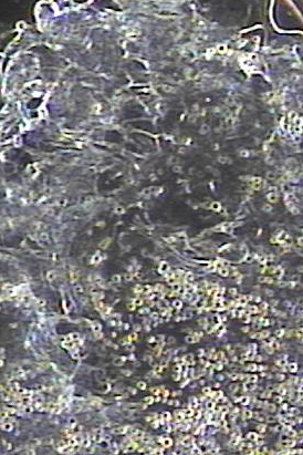
주 2회 전립선의 표적치료중 배출된 혈낭종과 사정관과 전립선관내 막혀있던 상피세포 덩어리 들의 현미경학적 관찰입니다.
Microscopic findings reveal hemorrhagic cysts and aggregated exfoliated epithelial cells that were previously obstructing the ejaculatory ducts and prostatic ducts, discharged during biweekly targeted prostate therapy.
주 2회 전립선의 4개월 표적 치료후 전립선의 결절이 감소하고 전립선내 다발성 혈낭종이 치료되고
혈낭종의 크기가 감소하고 있는 졍면 경직장 초음파사진입니다.
This is a frontal transrectal ultrasound image taken after four months of biweekly targeted prostate therapy, showing a reduction in prostatic nodules and multiple hemorrhagic cysts within the prostate, with a notable decrease in the size of the cysts.
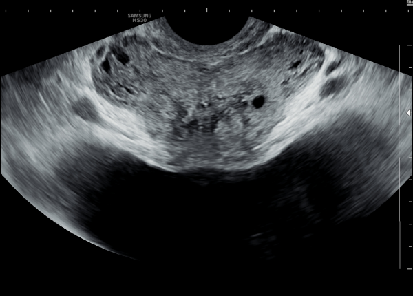
주 2회 전립선의 표적 치료후 다발성 낭종이 치료되고 있는 경직장 전립선 초음파 사진
"A transrectal prostate ultrasound image showing the treatment of multiple cysts after targeted prostate therapy twice a week."
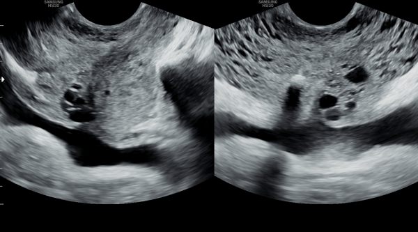
4개월 동안 주 2회 전립선과 정낭의 표적 치료후 다발성 정낭의 낭종들이 치료 되고 있는 경직장 전립선의 초음파 사진입니다.
This is a transrectal ultrasound image showing the improvement of multiple seminal vesicle cysts following four months of biweekly targeted therapy to the prostate and seminal vesicles.
주 2회 전립선과 사정관, 정낭 그리고 정관의 표적 치료후 고환의 섬유화가 감소하고 정상 크기의 고환의 초음파 사진입니다.
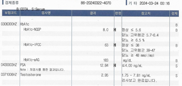
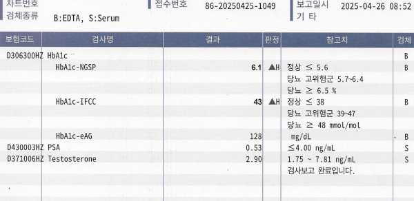
첫 내원 당일 혈액학적 검사상 당화 혈색소가 8.0 %, PSA 12.84 ng/mL, Testosterone 2.95 ng/mL 높은 수치를 보였습니다.
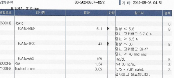
주 2회 전립선의 표적 치료후 추적 혈액학적 검사상 당화 혈색소가 6.1 %, PSA 1.54 ng/mL, Testosterone 3.06 ng/mL로 정상 수치를 보였습니다.
만성 전립선염 전립선 비대증 만성 골반통 증후군 전립선 암등의 전립선의 표적치료를 하면 남성 호르몬의 자연 증가와 PSA, 전립선암, 전립선 비대증으로 증가되는 PSA 수치의 감소 그리고 당뇨질환으로 고생하시는 분의 4개월 동안 주2회 전립선의 표적치료로 치료된 당화 혈색소의 감소등이 탁월한 결과지 입니다.???? 그러므로 건강한 삶을 유지키 위해 반드시 전립선의 표적치료를 하러가야합니다 이따가 뵙겠습니다 서울가정의학과의원 드림 ????
댓글목록
등록된 댓글이 없습니다.


