전립선 자료실
페이지 정보
본문
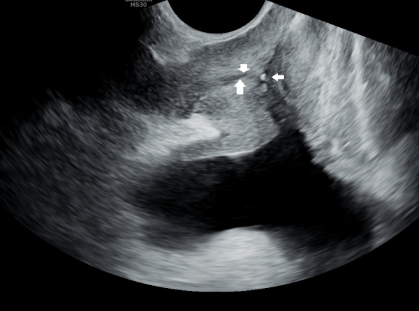
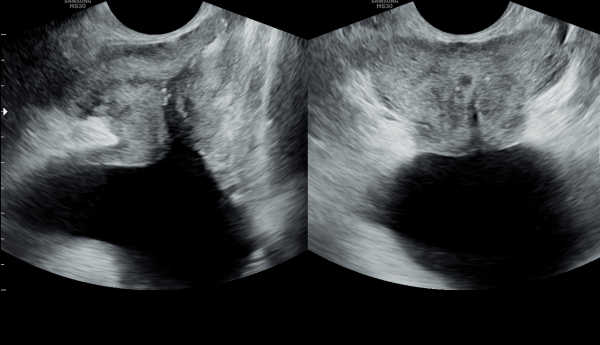
회음부 통증과 두통 그리고 오한이 있다고 내원당이 검사 한 경직장 전립선의 초음파 감사상 사정관 입구에 결석과 사정관 주위에 탈락된 상피 세포가 관찰되는 측면 전립선 초음파 사진.
This is a lateral transrectal prostate ultrasound image taken on the day of the patient's visit due to perineal pain, headache, and chills, showing calcifications at the ejaculatory duct opening and detached epithelial cells around the ejaculatory duct.
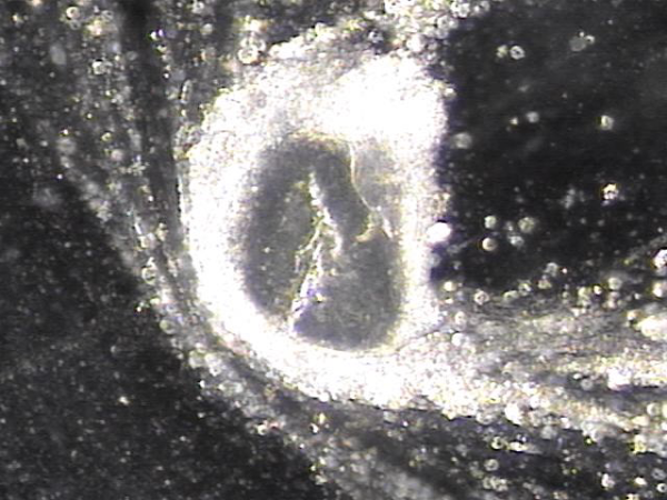
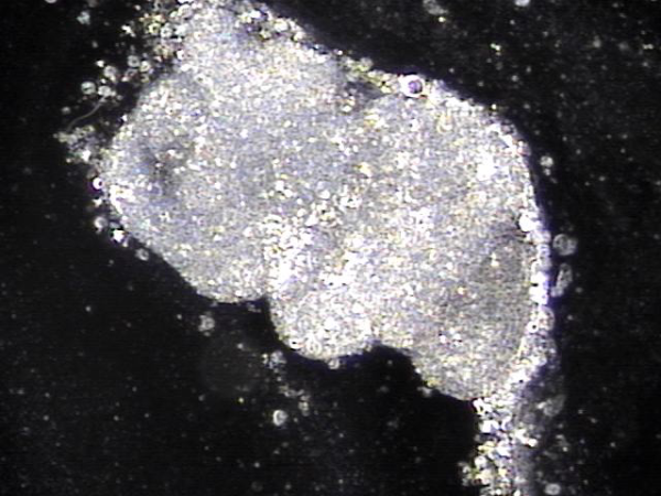
전립선의 표적 치료후 사정관과 정관에 막혀 있던 염증세포와 순환 장애를 일으키는 노페물의 현미경학적 검사 자료입니다.
This is a microscopic examination of inflammatory cells and waste materials causing circulatory obstruction that were blocked in the ejaculatory duct and vas deferens after targeted prostate treatment.
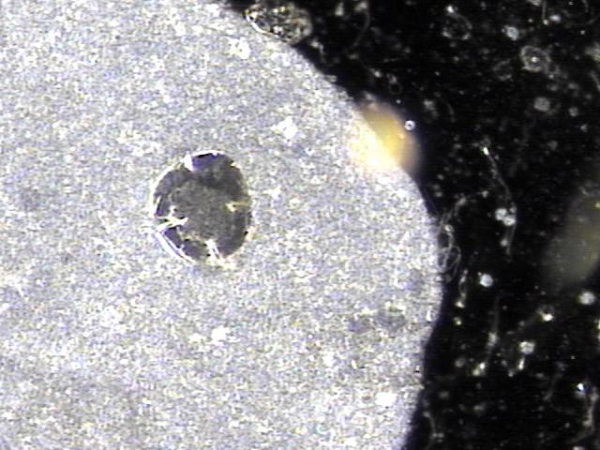
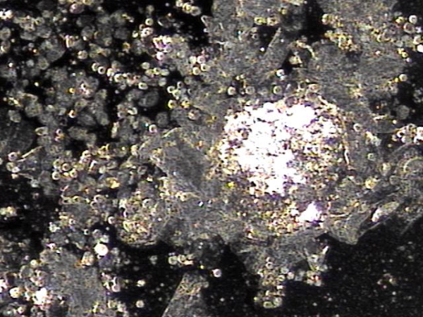
주 2회 전립선의 표적 치료중 배출된 사정관 입구의 결석과 정낭내 고여 있던 혈정액과 배출되지 않았던 노폐물들의 치료후 현미경학적 자료입니다.
This is a microscopic examination of the calculi discharged from the ejaculatory duct opening, along with the hemospermia and accumulated waste materials retained in the seminal vesicles, during twice-weekly targeted prostate treatment.
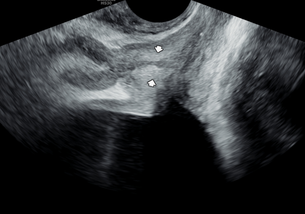
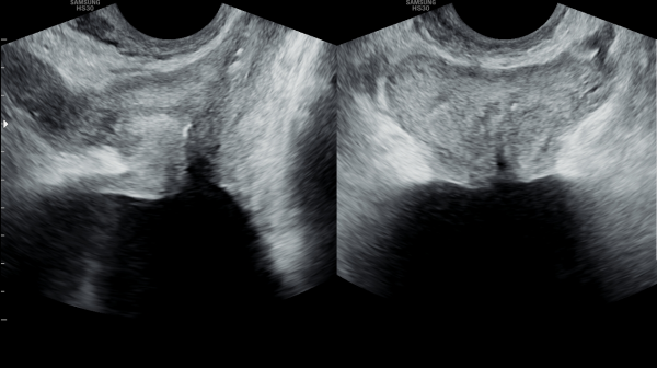
주 2회 전립선의 표적 치료 6개월뒤 사정관 입구의 결석이 치료되고 사정관 내에 막혀 있던 탈락된 상피 세포가 치료되어 사정관내 이물이 없어진 측면 경직장 전립선 초음파 추적 검사 자료입니다.
This is a follow-up lateral transrectal prostate ultrasound image taken six months after twice-weekly targeted prostate treatment, showing that the calculi at the ejaculatory duct opening have been treated and the previously obstructing desquamated epithelial cells within the ejaculatory duct have been resolved, leaving no foreign material inside the duct.
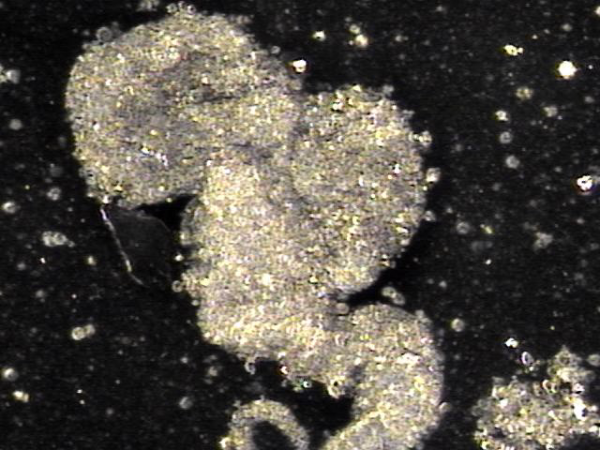
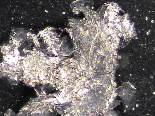
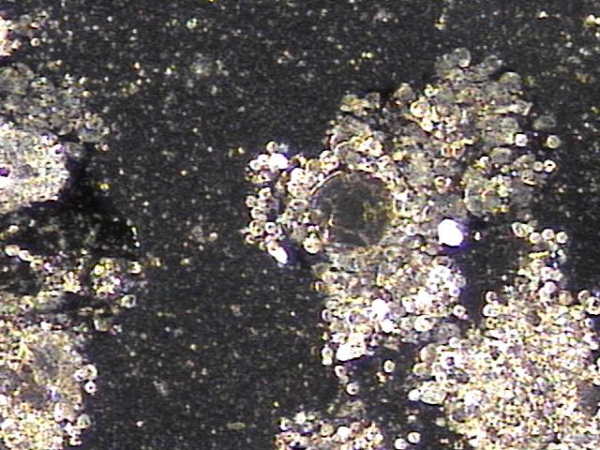
주 2회 전립선의 표적 치료후 배출된 염증 세포 덩어리와 결석의 현미경 학적 자료 입니다.
This is a microscopic examination of clumps of inflammatory cells and calculi discharged after twice-weekly targeted prostate treatment.
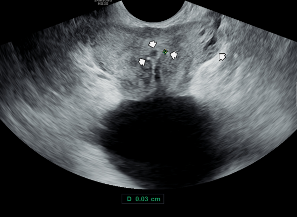
회음부 통증과 두통 그리고 오한이 있다고 내원당이 검사 한 경직장 전립선의 초음파 감사상 사정관 입구에 결석과 사정관 주위에 탈락된 상피 세포가 관찰되는 정면 전립선 초음파 사진상 좌측 사정관의 탈락된 상피세포로 사정관이 좁아지고(직경:0.03 Cm) 우측 사정관 입구의 미세 결석이 관찰되고 사정관 입구가 확장된 초음파 사진입니다.
On the day of the visit, due to perineal pain, headache, and chills, a transrectal prostate ultrasound was performed. The frontal ultrasound image shows epithelial cell debris around the ejaculatory ducts and a calculus at the duct opening. The left ejaculatory duct appears narrowed (diameter: 0.03 cm) due to accumulated epithelial debris, while a micro-calcification is observed at the opening of the right ejaculatory duct, which appears dilated.
주 2회 전립선의 표적 치료후 사정관과 정관 그리고 전립선관 등에서 배출된 상피세포 덩어리의 현미경학적 사진입니다.
This is a microscopic image of epithelial cell clumps discharged from the ejaculatory ducts, vas deferens, and prostatic ducts following targeted prostate treatment twice a week.
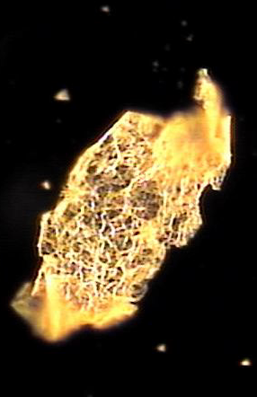
주 2회 전립선의 표적 치료후 사정관과 정관 그리고 전립선관 등에서 배출된 상피세포 덩어리의 현미경학적 사진입니다.
This is a microscopic image of epithelial cell clumps discharged from the ejaculatory ducts, vas deferens, and prostatic ducts following targeted prostate treatment twice a week.
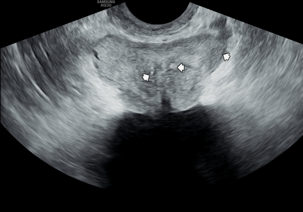
6개월간 주 2회 전립선의 표적 치료후 좌측 사정관의 직경이 회복되고 우측 사정관 입구를 막고 있는 결석의 크기가 줄어들고 좌측 전립선 결절이 감소하고 사정관 낭종이 치료된 추적 정면 경직장 전립선 초음파 사진입니다.
This is a follow-up frontal transrectal prostate ultrasound image after six months of targeted prostate treatment twice a week, showing recovery of the left ejaculatory duct diameter, reduction in the size of the stone blocking the right ejaculatory duct opening, decreased nodules in the left prostate, and resolution of ejaculatory duct cysts.
첫 내원 당일 검사한 경직장 전립선 초음파 사진중 측면 사진상 사정관내 탈락된 상피 세포가 쌓여 있고 사정관 입구에 결석이 있으며 정면 사진상 사정관 낭종과 사정관 입구내 결석이 관찰되고 있는 사진입니다.
On the first visit, transrectal prostate ultrasound images showed, in the lateral view, accumulation of desquamated epithelial cells within the ejaculatory duct and a stone at the ejaculatory duct opening; in the frontal view, an ejaculatory duct cyst and a stone within the ejaculatory duct opening were observed.
6개월동안 전립선의 표적 치료후 사정관의 낭종의 크기가 감소하고 좌측 사정관입구의 결석의 크기가 줄고 사정관내 쌓인 탈락된 상피 세포가 치료되고 전립선의 이행구역의 결절의 크기가 줄어든 추적 경직장 전립선의
초음파 사진입니다.
This is a follow-up transrectal prostate ultrasound image after six months of targeted prostate therapy, showing a reduction in the size of the ejaculatory duct cyst, a decrease in the size of the stone at the left ejaculatory duct opening, resolution of accumulated desquamated epithelial cells within the ejaculatory duct, and a decrease in the size of a nodule in the transitional zone of the prostate.
주 2회 사정관 입구의 표적 치료중 치료된 혈정액등의 현미경학적 자료입니다.
This is a microscopic image of treated hemospermia obtained during targeted therapy of the ejaculatory duct opening performed twice a week.
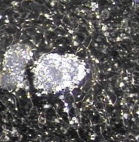
주2회 전립선의 표적 치료중 정관과 사정관 등에서 배출된 현미경학적 자료입니다.
Microscopic findings of materials discharged from the vas deferens and ejaculatory ducts during twice-weekly targeted prostate treatment.
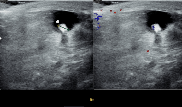
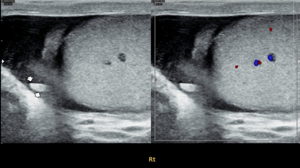
첫내원 당일 고환과 부고환의 초음파 검사상 결석이 관찰되는 검사 자료입니다.
This is an ultrasound image taken on the first visit, showing the presence of calcifications in the testis and epididymis.
주2회 전립선의 표적 치료중 정관과 사정관 등에서 배출된 현미경학적 자료입니다.
Microscopic findings of materials discharged from the vas deferens and ejaculatory ducts during twice-weekly targeted prostate treatment.
주2회 전립선의 표적 치료중 정관과 사정관 등에서 배출된 현미경학적 자료입니다.
Microscopic findings of materials discharged from the vas deferens and ejaculatory ducts during twice-weekly targeted prostate treatment.
6개월간 주 2회 전립선과 사정관, 정낭 그리고 정관의 표적 치료후 고환과 부고환의 초음파 검사상 결석의 크기가 감소하고 있는 추적 경직장 전립선의 초음파 자료입니다.
Follow-up transrectal prostate ultrasound findings after six months of twice-weekly targeted treatment of the prostate, ejaculatory ducts, seminal vesicles, and vas deferens, showing a reduction in the size of calcifications in the testes and epididymis.
모든 남성은 누구던지 반드시 기억해야 할 의학적인 지식은 매일 신체의 각 부위에서 충실히 역활을 한 세포는 1~3주내 수명을 다하고 탈락하며 새로운 상피세포가 만들어져 각 장기에 그 역활을 이어가나 전립선의 상피세포는 탈락하여 좁고 긴 배출관(직경 : 0.1~0.2mm)에 쌓여 비대해 지고 낭종과 섬유화와 결석등이 진행하여 여러 증상이 생겨 온갖 광고에 유혹 되기도 하고 잘못된 치료의 길도 갑니다.
꼭 기억 해야할 치료는 오랫동안 쌓인 상피세포 덩어리는 수년간~ 반복 표적치료로 휴유증 없이 치료된다는 자료입니다. 포기하지 마라 포기하지 마라 결코 포기하지 마라 라고 외친 윈스턴 처칠경의 말씀을 새기면서 그리고 순환과 지속적인 관리를 위해 반드시 전립선의 표적치료를 하러가야합니다.
이따가 뵙겠습니다.
An essential piece of medical knowledge that every man must remember is that epithelial cells throughout the body, after fulfilling their roles, naturally shed within 1 to 3 weeks and are replaced by new cells to maintain organ function. However, in the prostate, the desquamated epithelial cells tend to accumulate in the narrow and elongated ducts (diameter: 0.1–0.2 mm), leading to prostate enlargement, cyst formation, fibrosis, and calcifications, which cause various symptoms. This can make men susceptible to misleading advertisements and ineffective treatments.
What must be remembered is that even epithelial cell masses accumulated over years can be successfully treated without sequelae through repeated, targeted therapy over time. Let us take to heart the words of Sir Winston Churchill: 'Never give up, never give up, never, never, never give up.' And for proper circulation and ongoing care, targeted prostate treatment is essential.
See you later.
- 다음글전립선 자료실 25.04.18
댓글목록
등록된 댓글이 없습니다.


