전립선자료실
페이지 정보
본문
주2회 전립선의 표적 치료전 정면 경직장 전립선 초음파 사진상 좌우측 사정관 입구의 결석과 우측 전립선내 결석이 관찰되는 초음파 사진입니다.
This is a frontal transrectal prostate ultrasound image taken before twice-weekly targeted prostate treatment, showing calculi at the openings of both ejaculatory ducts and a calculus within the right side of the prostate.
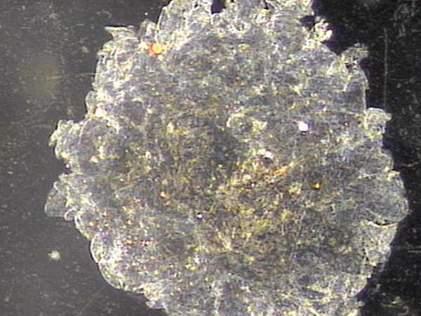
주 2회 전립선의 표적 치료중 탈락된 상피 세포가 사정관과 정관 그리고 전립선관에서 배출된 현미경학적 사진입니다.
This is a microscopic image showing exfoliated epithelial cells discharged from the ejaculatory duct, vas deferens
and prostatic duct during twice-weekly targeted prostate treatment.
주 2회 전립선의 표적 치료후 양측 사정관 입구의 결석과 우측 전립선내 결석이 감소하고 있는 추적 정면 경직장 전립선 초음파 사진입니다.
This is a follow-up frontal transrectal ultrasound image of the prostate showing a reduction in calculi at the bilateral ejaculatory duct openings and in the right side of the prostate after twice-weekly targeted prostate treatment.
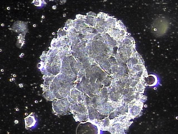
전립선의 표적 치료중 탈락되어 쌓여 있던 상피세포 덩어리의 현미경학적 사진입니다.
This is a microscopic image of accumulated epithelial cell clumps that were shed during targeted prostate treatment.
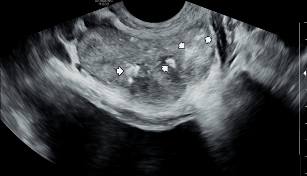
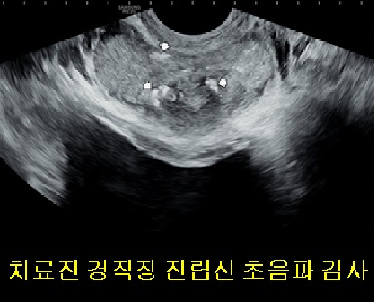
서울가정의학과의원에 내원 당일 경직장 전립선 초음파 검사상 양쪽 사정관 입구의 결석과 좌측 전립선의 중심 구역에 생긴 결석이 관찰된 자료입니다.
"This is the data from the day of the visit to the Seoul Family Medicine Clinic, showing the presence of stones at the entrances of both ejaculatory ducts and a stone located in the central zone of the left prostate, as observed on the transrectal prostate ultrasound."
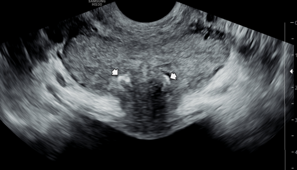
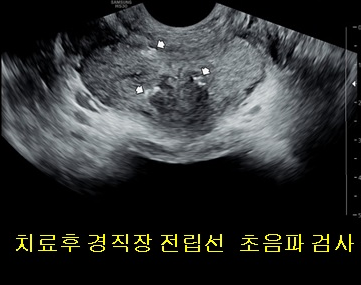
주2회 전립선의 표적치료후 양측 사정관 입구의 결석과 좌측 전립선의 중심 구역에 생긴 결석이 치료 되고 있는 경직장 전립선 초음파 사진
"This is a transrectal prostate ultrasound image showing the treatment of stones at the entrances of both ejaculatory ducts and a stone in the central zone of the left prostate, following targeted prostate therapy twice a week."
댓글목록
등록된 댓글이 없습니다.


